
A cranial nerve examination is crucial for diagnosing neurological conditions, assessing the 12 nerves originating from the brain, which control functions like smell, vision, hearing, and muscle movement.
Importance of Cranial Nerve Examination
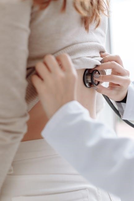
A cranial nerve examination is essential for identifying abnormalities in the 12 cranial nerves, which control critical functions like vision, hearing, smell, and motor skills. It helps diagnose conditions affecting the brainstem, cranial nerve pathways, or peripheral nerves. Early detection of deficits can lead to timely interventions, improving patient outcomes. The examination is non-invasive and provides valuable insights into neurological health. It is particularly vital for patients with symptoms like vision loss, facial weakness, or swallowing difficulties. By assessing each nerve’s function, clinicians can pinpoint lesions or impairments, guiding further investigations and treatment plans. This targeted approach ensures comprehensive neurological care, making cranial nerve examinations a cornerstone of clinical practice.
Overview of Cranial Nerves
The cranial nerves are 12 pairs of nerves originating from the brain, each responsible for specific sensory, motor, or parasympathetic functions. They are designated by Roman numerals I to XII and named according to their primary roles. For instance, the olfactory nerve (I) manages smell, while the optic nerve (II) handles vision. The oculomotor (III), trochlear (IV), and abducens (VI) nerves control eye movements, and the trigeminal (V) and facial (VII) nerves manage facial sensation and expression. The vestibulocochlear nerve (VIII) is vital for hearing and balance. The glossopharyngeal (IX), vagus (X), accessory (XI), and hypoglossal (XII) nerves regulate swallowing, voice, neck movements, and tongue function. Understanding their roles is key to diagnosing and managing neurological disorders effectively.

Overview of Cranial Nerves
The 12 cranial nerves originate from the brain, controlling vital functions like sensation, movement, and autonomic responses. Their examination is key to identifying neurological deficits and diagnosing conditions.
Listing the 12 Cranial Nerves
The 12 cranial nerves are essential for various bodily functions, each with distinct roles. They include:
- I (Olfactory) ─ responsible for smell.
- II (Optic) — governs vision.
- III (Oculomotor) ─ controls eye movement and pupil response.
- IV (Trochlear) — manages superior oblique eye muscle function.
- V (Trigeminal) — handles facial sensation and chewing.
- VI (Abducens) ─ controls lateral eye movement.
- VII (Facial) ─ regulates facial expressions and taste.
- VIII (Vestibulocochlear) — manages hearing and balance.
- IX (Glossopharyngeal), involved in taste, swallowing, and salivation.
- X (Vagus) ─ controls various functions like heart rate and digestion.
- XI (Accessory) — governs neck and shoulder muscle movement.
- XII (Hypoglossal) — regulates tongue movement for speech and swallowing.
These nerves are fundamental to diagnosing neurological conditions and understanding the brain’s functional connections.
Functions and Significance
The 12 cranial nerves perform distinct yet interconnected functions vital for sensory and motor activities. They regulate essential processes such as vision, hearing, smell, taste, and swallowing, as well as facial expressions and eye movements. Cranial nerves also control autonomic functions like heart rate and digestion. Their significance lies in their role as pathways for transmitting signals between the brain and peripheral structures, enabling precise communication and coordination. Damage to these nerves can lead to severe impairments, making their examination critical for diagnosing neurological disorders. Understanding their functions aids in identifying deficits, guiding targeted treatments, and improving patient outcomes. This underscores the importance of a thorough cranial nerve examination in clinical practice.
Preparation for the Examination
Ensure patient consent, proper positioning, and a quiet environment. The examiner should introduce themselves, explain the process, and gather necessary equipment for a thorough assessment.
Patient Preparation
Patient preparation is essential for an effective cranial nerve examination. The patient should be seated comfortably in a chair, positioned at eye level with the examiner. Ensure the room is well-lit to facilitate accurate visual assessments. Remove any hearing aids or glasses that may interfere with specific tests. The patient should be informed about the examination process to reduce anxiety and ensure cooperation. Avoid strong odors that could interfere with olfactory testing. The patient should also be asked about any pre-existing conditions that may impact the examination, such as vision or hearing impairments. Proper preparation ensures a smooth and accurate assessment of cranial nerve function.
Examiner Preparation
Examiner preparation is vital for a thorough cranial nerve examination. Gather necessary equipment, such as a tuning fork for hearing assessments, olfactory kits for smell testing, a penlight for pupil reactions, and gloves for patient interaction. Review the patient’s medical history to identify potential deficits or conditions that may impact the exam. Ensure the examination room is well-lit and quiet to minimize distractions. Introduce yourself to the patient, explain the process, and obtain consent. Wash your hands and maintain proper hygiene throughout the examination. Organize the examination systematically, testing each cranial nerve in sequence to avoid missing any. Proper preparation ensures accuracy and efficiency in assessing cranial nerve function.
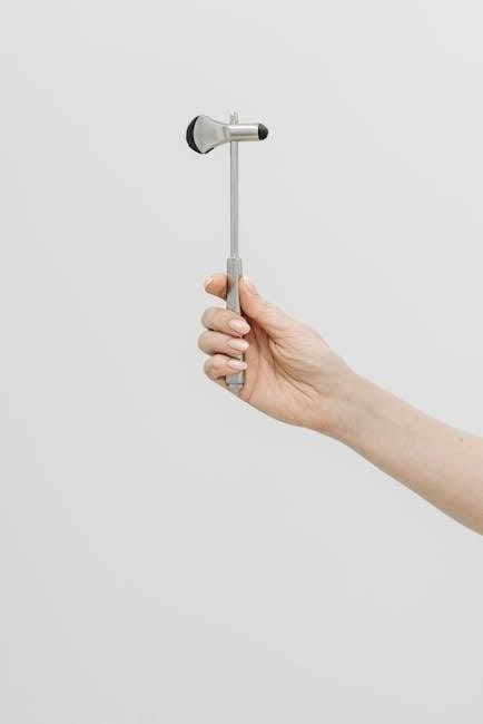
Equipment Needed
The equipment required for a cranial nerve examination includes a penlight for assessing pupil reactions, a tuning fork for hearing tests, and an olfactory kit with familiar scents for smell assessment. A reflex hammer is used to evaluate motor responses, and gloves are essential for patient interaction. Additional tools include a tongue blade for gag reflex testing and a mirror for observing uvula movement. Ensure all items are readily available to streamline the examination process and maintain patient comfort. Proper equipment preparation is key to accurately assessing cranial nerve function and identifying potential abnormalities.
Examination Process
The cranial nerve examination systematically evaluates each nerve’s function, starting with the olfactory nerve and progressing through to the hypoglossal nerve, ensuring thorough assessment of sensory and motor responses.
Cranial Nerve I (Olfactory)
Cranial Nerve I, the olfactory nerve, is responsible for transmitting sensory information related to smell. To assess its function, the patient is asked if they have noticed any changes in their sense of smell. The examiner then uses standardized odor bottles, such as coffee or peppermint, to test each nostril separately. The patient should identify the scent with each nostril tested individually. This evaluation helps detect abnormalities such as anosmia (loss of smell) or hyposmia (reduced olfactory sensation). The olfactory nerve examination is crucial for identifying potential neurological conditions, including those affecting the frontal lobe or olfactory pathways. It is often the first step in a comprehensive cranial nerve assessment, providing insights into sensory function and overall neurological health.
Cranial Nerve II (Optic)
Cranial Nerve II, the optic nerve, is responsible for vision and transmitting visual information from the retina to the brain. Assessment begins with inspecting the pupils for symmetry, reactivity to light, and accommodation. Visual acuity is tested using a Snellen chart or near vision cards. The examiner evaluates extraocular movements to ensure proper eye alignment and tracking. Direct and consensual pupillary reflexes are checked to confirm intact afferent pathways. Fundoscopic examination Using an ophthalmoscope allows visualization of the optic disc, retina, and macula, helping identify abnormalities such as papilledema or optic atrophy. Any deviation in pupil responses or visual field defects may indicate optic nerve dysfunction, potentially signaling conditions like optic neuritis or tumors. This nerve’s examination is critical for diagnosing visual impairments and neurological disorders affecting the optic pathways.
Cranial Nerve III (Oculomotor)
Cranial Nerve III, the oculomotor nerve, controls most eye movements, pupil constriction, and eyelid elevation. Examination involves assessing extraocular movements by asking the patient to follow the examiner’s finger in all directions. Pupillary reflexes are tested by shining a light into each eye to observe constriction. The examiner also checks for ptosis (drooping eyelid) and evaluates the ability to open the eye against resistance. Abnormal findings, such as limited eye movement, a dilated pupil, or ptosis, may indicate nerve dysfunction. This nerve is critical for eye function, and its impairment can suggest conditions like aneurysms, ischemia, or neurological disorders affecting the oculomotor pathways. Accurate assessment ensures early detection of potential abnormalities.
Cranial Nerve IV (Trochlear)
Cranial Nerve IV, the trochlear nerve, is responsible for innervating the superior oblique muscle, which controls downward and inward eye movements. During examination, the patient is asked to look downward while moving their eyes medially. Weakness or paralysis of this nerve may result in difficulty moving the eye in this direction, leading to diplopia or compensatory head tilting. The trochlear nerve has the longest intracranial course, making it susceptible to injury from trauma or conditions like multiple sclerosis. Testing this nerve involves observing the patient’s ability to perform precise eye movements and ensuring the absence of nystagmus or other ocular motor deficits. Accurate assessment is vital for diagnosing lesions affecting this nerve and guiding appropriate treatment.
Cranial Nerve V (Trigeminal)
Cranial Nerve V, the trigeminal nerve, is primarily responsible for sensory functions of the face and motor control of the muscles of mastication. It has three main divisions: the ophthalmic, maxillary, and mandibular branches; During examination, sensory function is tested by lightly touching the face with a soft object or assessing pain perception with a sharp object. Motor function is evaluated by observing the contraction of the masseter and temporalis muscles during jaw clenching. The corneal reflex, involving the ophthalmic and facial nerves, is also assessed. Abnormalities may include sensory deficits, muscle weakness, or reduced reflexes, which could indicate conditions such as trigeminal neuralgia or nerve compression. Accurate testing is essential for diagnosing and managing related neurological or facial disorders.
Cranial Nerve VI (Abducens)
Cranial Nerve VI, the abducens nerve, is responsible for controlling the lateral rectus muscle, which governs eye abduction. During the examination, the patient is asked to move their eyes laterally while the examiner observes for smooth, coordinated movement. Weakness or paralysis of the abducens nerve may result in medial strabismus (esotropia) or difficulty moving the affected eye outward. The nerve’s long intracranial course makes it susceptible to damage from conditions like increased intracranial pressure, tumors, or stroke. Testing the abducens nerve is critical for identifying cranial nerve palsies and assessing brainstem function. Abnormal findings may include nystagmus or limited eye movement, necessitating further neurological evaluation to determine the underlying cause.
Cranial Nerve VII (Facial)
Cranial Nerve VII, the facial nerve, controls facial expressions, taste sensation on the anterior tongue, and some autonomic functions; During the examination, the patient is asked to perform movements such as smiling, frowning, and showing teeth to assess symmetry and strength of facial muscles. The examiner also tests taste by applying sweet, sour, salty, or bitter substances to the anterior tongue. Weakness or paralysis, such as Bell’s palsy, may result in facial droop or asymmetry; The nerve’s sensory and motor functions are critical for identifying lesions, which can occur from conditions like strokes, tumors, or infections. Abnormal findings, such as decreased taste or facial weakness, can indicate neurological issues requiring further investigation. Accurate assessment of CN VII is essential for diagnosing cranial nerve palsies and underlying pathologies.
Cranial Nerve VIII (Vestibulocochlear)
Cranial Nerve VIII, the vestibulocochlear nerve, is responsible for hearing and balance. The examination involves assessing both the cochlear (hearing) and vestibular (balance) components. For hearing, tests such as the Weber and Rinne tests are used. The Weber test involves placing a vibrating tuning fork on the forehead to assess sound lateralization, while the Rinne test compares bone conduction to air conduction. For vestibular function, the examiner may perform the Dix-Hallpike maneuver to check for nystagmus or vertigo. Abnormal findings, such as unilateral hearing loss or vestibular dysfunction, may indicate conditions like labyrinthitis, Ménière’s disease, or acoustic neuromas. Accurate evaluation of CN VIII is critical for diagnosing auditory and vestibular pathologies, ensuring proper management of hearing and balance disorders.
Cranial Nerve IX (Glossopharyngeal)
Cranial Nerve IX, the glossopharyngeal nerve, is examined to assess its motor and sensory functions, including swallowing, taste, and salivation. The gag reflex is tested by gently stimulating the pharyngeal wall with a tongue depressor, observing for symmetric contraction of the pharyngeal muscles. The patient is asked to say “ahh” to inspect the uvula and soft palate for elevation and symmetry. Asymmetric movement or a diminished gag reflex may indicate nerve dysfunction. This nerve also supplies the parotid gland, and its sensory role in taste can be assessed by applying a taste stimulus to the posterior tongue. Abnormal findings may suggest lesions affecting the glossopharyngeal nerve, potentially impacting swallowing and oral functions.
Cranial Nerve X (Vagus)
Cranial Nerve X, the vagus nerve, is assessed for its extensive functions, including motor, sensory, and parasympathetic roles. The motor component is evaluated by observing the patient’s ability to perform tasks like coughing and speaking, as well as the movement of the soft palate and uvula during phonation. The gag reflex, tested by stimulating the pharynx, also involves the vagus nerve. Sensory function is examined by testing taste on the epiglottis. Additionally, the patient’s ability to swallow is assessed, as the vagus nerve controls the pharyngeal muscles. Abnormal findings, such as hoarseness, dysphagia, or a diminished gag reflex, may indicate vagal nerve dysfunction, which can be associated with various conditions, including stroke or peripheral neuropathy.
Cranial Nerve XI (Accessory)
Cranial Nerve XI, the accessory nerve, is primarily responsible for controlling the sternocleidomastoid and trapezius muscles, which are essential for shoulder movement and head rotation. During the examination, the patient is asked to shrug their shoulders and turn their head against resistance to assess motor function. The examiner evaluates the strength and symmetry of these movements. Weakness or asymmetry may indicate nerve damage or dysfunction. The accessory nerve is unique as it has both cranial and spinal roots, with the cranial portion contributing to swallowing and vocal cord function via the vagus nerve. Abnormalities, such as atrophy or reduced muscle tone, can suggest conditions like traumatic injury, neurological disorders, or congenital defects. Accurate assessment of CN XI is vital for diagnosing motor impairments affecting the neck and shoulders.
Cranial Nerve XII (Hypoglossal)
Cranial Nerve XII, the hypoglossal nerve, governs tongue movements, including protrusion, lateral deviation, and anterior-posterior motion. During examination, the patient is asked to protrude their tongue and move it side-to-side. The examiner observes for atrophy, fasciculations, or deviation, which may indicate nerve damage. Weakness or paralysis can lead to difficulty in speech and swallowing. The hypoglossal nerve’s unilateral lesion causes the tongue to deviate toward the affected side upon protrusion. This nerve is crucial for assessing brainstem integrity and identifying conditions like stroke or neurodegenerative diseases. Accurate evaluation ensures proper diagnosis and management of related motor and swallowing disorders.

Common Abnormalities and Interpretation
Cranial nerve abnormalities often manifest as weakness, paralysis, or sensory deficits. These findings help identify nerve-specific lesions, guiding diagnosis and treatment of underlying conditions like strokes or neuropathies.
Recognizing Abnormal Findings
Abnormal findings in a cranial nerve exam may include motor deficits like weakness or paralysis, sensory deficits such as numbness, or reflex abnormalities. For example, ptosis, diplopia, or nystagmus may indicate issues with cranial nerves III, IV, or VI. Facial asymmetry or difficulty smiling could suggest a problem with cranial nerve VII. Hearing loss or vertigo may point to cranial nerve VIII dysfunction. Weakness in tongue movement or speech difficulties could signal issues with cranial nerves IX, X, or XII. Proper documentation of these findings is essential for accurate diagnosis and treatment planning. Correlating these abnormalities with patient history and additional tests like imaging or lab work helps pinpoint the underlying cause, whether it’s a lesion, infection, or neurological disorder.
Interpreting Test Results
Interpreting cranial nerve test results involves correlating findings with patient symptoms and medical history. Normal results indicate intact cranial nerve function, while abnormalities suggest potential lesions or neurological conditions. For instance, unilateral hearing loss may indicate a vestibulocochlear nerve issue, while facial weakness could point to a facial nerve lesion. Oculomotor deficits, such as ptosis or diplopia, may suggest brainstem or cranial nerve pathology. Abnormal reflexes, like a diminished gag reflex, could indicate glossopharyngeal or vagus nerve dysfunction. Accurate interpretation requires understanding each nerve’s function and possible causes of dysfunction, such as trauma, infections, or systemic diseases. Comprehensive documentation and further diagnostic testing, like MRI or EMG, may be necessary to confirm the diagnosis and guide appropriate management. Timely interpretation ensures effective patient care and treatment planning.

Case Studies and Clinical Applications
Clinical applications of cranial nerve examinations are evident in diagnosing conditions like multiple cranial nerve palsies and oculomotor nerve palsy. Real-world examples, such as a woman with IIH-related oculomotor nerve palsy, illustrate practical relevance.
Real-World Examples
Real-world examples highlight the practical application of cranial nerve examinations in diagnosing neurological conditions. For instance, a woman in her 50s presented with IIH-related oculomotor nerve palsy, showing dilated pupils, ptosis, and medial rectus palsy. This case underscores the importance of cranial nerve assessments in identifying specific nerve involvement. Similarly, a case series of 14 Thai patients with recurrent multiple cranial nerve palsies demonstrated how examinations can reveal patterns of nerve dysfunction. Another example involved a solitary mass near the trigeminal nerve, causing facial pain and sensory loss, which was diagnosed through targeted cranial nerve testing. These scenarios illustrate how cranial nerve exams are essential for localizing lesions and guiding clinical management, emphasizing their role in everyday neurological practice.
Clinical Relevance
Cranial nerve examinations hold significant clinical relevance as they provide critical insights into neurological function and dysfunction. By assessing the 12 cranial nerves, healthcare providers can identify specific nerve involvement, guiding diagnosis and treatment. For instance, abnormalities in the trigeminal nerve may indicate conditions like trigeminal neuralgia, while oculomotor nerve palsy can signal increased intracranial pressure. These examinations are essential for localizing lesions, monitoring disease progression, and evaluating recovery. Clinicians rely on cranial nerve tests to detect conditions such as Bell’s palsy, multiple sclerosis, or stroke, where nerve dysfunction is a hallmark. Early detection of abnormalities can lead to timely interventions, improving patient outcomes. Thus, cranial nerve exams are a cornerstone of neurological practice, enabling precise and targeted care.
A cranial nerve examination is a fundamental tool in neurology, offering insights into nervous system function and aiding in the diagnosis of various neurological disorders effectively.
A cranial nerve examination is essential for assessing the function of the 12 cranial nerves, which control vital functions like sensation, movement, and involuntary processes. Proper preparation, including patient positioning and equipment readiness, ensures accurate results. Each nerve has specific tests, such as assessing smell for CN I or checking eye movements for CN III and IV. Abnormal findings, like weakness or sensory deficits, indicate potential neurological issues. Accurate documentation and interpretation of results are critical for diagnosis and treatment planning. This examination is a cornerstone in neurology, aiding in the early detection and management of conditions affecting the brain and nervous system. By following a structured approach, healthcare professionals can effectively evaluate cranial nerve function and provide tailored care.
Documentation Best Practices
Accurate and detailed documentation is critical for cranial nerve examinations. Use standardized templates to record findings systematically, ensuring clarity and consistency. Note any abnormalities, such as weakness, sensory deficits, or reflex changes, using precise terminology. Include the date, time, and patient identifiers for accountability. Document both positive and negative findings to provide a complete picture; Organize results in a logical sequence, aligning with the examination process. Use electronic health records (EHRs) for easy access and secure storage. Ensure confidentiality by adhering to HIPAA guidelines and institutional policies. Clear documentation aids in tracking patient progress, facilitates communication among healthcare providers, and supports legal and academic purposes. Regularly review and update records to reflect ongoing patient care and outcomes.